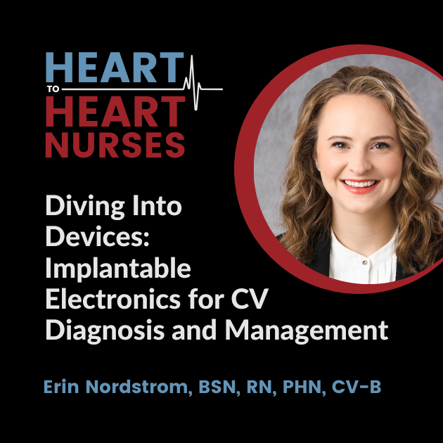
The wide array of cardiovascular implantable electronic devices (CIEDs) may lead to confusion as to which one to use in what circumstances. Differentiating defibrillators, pacemakers, and loop recorders–as well as accessing and utilizing the collected data–is covered by guest Erin Nordstrom, BSN, RN, PHN, CV-B.
Episode Resources
- Choosing the Right Cardiac Pacing Device - 0.75 CE contact hours
- 12-Lead ECG in the Clinical Setting - 2.0 CE contact hours
- Managment of Atrial Fibrillation - 0.65 CE contact hours, 0.2 CE pharmacology hours
Welcome to Heart to Heart Nurses, brought to you by the Preventive Cardiovascular Nurses Association. PCNA's mission is to promote nurses as leaders in cardiovascular disease prevention and management.
Geralyn Warfield (host): (00:20) Welcome to today's episode where we have the great, great privilege of talking with a nurse who's going to be speaking with us about implantable devices. I'd like Erin Nordstrom to introduce herself to you.
Erin Nordstrom (guest): (00:32) Sure, So, I currently work as a cardiac rhythm device nurse for Metropolitan Heart and Vascular. So, I work in a device clinic where we interrogate pacemakers and defibrillators for patients who have heart rhythm disorders
Geralyn Warfield (host): (00:48) And in that capacity, what kind of devices do you see in that setting?
Erin Nordstrom (guest): (00:55) So, there are a lot of different kinds of devices on the market, but we call them cardiovascular implantable electronic devices. So, we call them CIEDs for short, and these include pacemakers, defibrillators, and loop recorders.
So, pacemakers pace the heart, and they're typically implanted for those who have cardiac dysrhythmias that caused the heart rate to go too low. So, patients with different types of AV block or sick sinus syndrome, and that way their heart rate doesn't go too low—that's the purpose of a pacemaker. The purpose of a pacemaker is not to fix any fast type of heart rhythm, which is a common misconception that you'll find patients feel, but the only purpose is to prevent that heart rate from going too low.
There's also defibrillators. On the other hand, these are implanted to fix faster heart rhythms. And in a typical transvenous system where leads are implanted into the heart—right atrium, right ventricle—these can also pace and they sense fast heart rhythms. So, they can deliver therapy like ATP and shocks to get patients out of dangerous arrhythmias like ventricular fibrillation and ventricular tachycardia. And they're implanted for those who are at risk of sudden cardiac death. So, for either primary prevention or secondary prevention.
There are different defibrillators on the market. There was one defibrillator called subcutaneous defibrillator, which is implanted on a patient's left side in subcutaneous tissue, and a lead is run over the abdomen and up through the sternum. So those types of defibrillators are only there to deliver therapy, not necessarily pace if they need to.
That's also a common patient misconception is that if they have a defibrillator, it's only there to shock them or deliver types of therapy, but actually transvenous defibrillators have the ability to pace the heart as well.
Loop recorders are small devices that are implanted in subcutaneous tissue right in the middle of the chest. These are much less technical. We don't get as much data from these, but you can think of it as a long-term Holter monitor. So, they last about three to five years now. All device manufacturers make them. And they're great because we can use them to rule out types of...people who have syncope that's unexplained, for cryptogenic stroke patients to rule out AFib, and those who are having palpitations or symptoms that don't occur often enough for us to catch it on a type of Holter monitor.
Geralyn Warfield (host): (03:59) Well, thanks so much for going through that whole series of things that might be happening in your facility. And I'm wondering how patients are when they come to you. I suspect like all patients that are coming for some kind of procedure or even a clinic visit, there are some that are, you know, have done a ton of research and know exactly what's going on. And there are others that still aren't quite sure even when they arrive in your facility. Is that accurate?
Erin Nordstrom (guest): (04:28) Yes, yes. So, I think the majority of patients that we see are quite unfamiliar with these devices and how they function. So typically, after a device is implanted by an electrophysiologist, they will follow up with a device clinic within 7-10 days. And this is just our clinic, our protocol. And at that visit, we do a ton of patient education about the device, how it functions, why it was implanted, what kind of cardiac dysrhythmia they have and how this will fix them.
So, I should mention that there are devices that have three leads and those are implanted for those who have heart failure with a left bundle branch block. We do provide some heart failure education at that time.
And then we also provide the patient a home monitoring device. So, a patient's home monitor is a middleman between the clinic and a patient's device. So, we can actually pull data from that home monitor that the patient has. So, we'll give that to them at the clinic check.
In addition, we assess the patient's wound and take the bandages off and make sure there's no hematoma or signs of infection. And then we actually check the pacemaker, the defibrillator with a programmer. So that, again, is within 7-10 days after implant.
Then we'll pull a remote transmission in about a month after their implant. And then we see them again in clinic for manual testing three months after implant. From that point on, if everything looks stable—because it takes about at least four to six weeks for those leads to really embed into the cardiac tissue and heal and this fibrotic capsule forms—then after that three-month appointment, we can see them in clinic every one to two years.
And we pull remote transmissions every three months to ensure that the technical data looks good.
Geralyn Warfield (host): (06:25) And for those devices that have a limited shelf life, is that replacement happening in your facility as well?
Erin Nordstrom (guest): (06:31) Yes, yes. So we are alerted via the home monitor, which I should say as well, there's apps available now. We get alerted three months prior to when they reach recommended replacement time to ensure they have time to do a pre-op and get the device replaced. So, our facility does that as well.
Geralyn Warfield (host): (06:51) We've been talking about implantable devices, and we will take a quick break and be right back.
Geralyn Warfield (host): We'd like to welcome you back to our conversation with Erin Nordstrom. And Erin, there were a couple other pieces about implantable devices that you wanted to make sure that our audience heard.
Erin Nordstrom (guest): (07:06) Yeah. So, I wanted to talk about how these devices are actually implanted. So, when you think of a traditional pacemaker or defibrillator, a transvenous system, these are implanted into subcutaneous tissue right under the patient's clavicle. Sometimes, rarely, it's done subpectorally just based on the patient's anatomy. And the electrophysiologist can insert leads usually through the subclavian and/or the cephalic vein and then the leads are tunneled down into the heart's chambers.
So, there's different types of systems. So, you can have a device with one lead, two leads, or three. We call a device with one lead a single chamber device. With two leads it's a dual chamber device. And with that third left ventricular lead it's just a multi-pacing system.
So, that first lead could be implanted into the right atrium or the right ventricle. I think what we see most commonly now, at least where I am in Minnesota, is we're implanting more dual chamber devices because that way we can see what's going on in the top of the heart as well as the bottom of the heart. So, you'll have a lead in the right atrium and the right ventricle.
That third lead when they implant that, it's a little bit more difficult of an implant for the electrophysiologist because it can be based on the patient's anatomy. But that lead is put into a lateral vein on the left side of the heart. And again, the indication for heart failure in left bundle branch block, the idea is that we can then synchronize the left and the right side of the heart and improve one's injection fraction.
Geralyn Warfield (host): (08:52) We've talked a lot about what the devices are, how they're implanted, but I think the reason for having all these processes in place is to get that data out. Could you talk a little bit about how that process works?
Erin Nordstrom (guest): (09:03)
Yeah, so how a device is interrogated, a patient doesn't have to do anything. But again, when we see them in clinic, they'll sit down in a chair, And we bring out these big chunky programmers that look like very big computers. Each device manufacturer has a different programmer. And they come with a type of wand and this wand is placed over a patient's device and then we have connection.
So, during clinic testing, we can look through a ton of different data. A lot of it is more diagnostic and technical, so we can see what's going on with the leads and the generator. But what is more exciting now is we're getting a lot of more clinical data from these devices as well.
Just running through a basic test, basic testing that we do at each clinic visit, we do three different types of tests.
One test is called impedance, and this is a measurement of the integrity of the lead. So, if there is a tear in the silicone or polyurethane covering of that lead, we would know. The second test we do is called sensing, and this is where we purposely lower a patient's heart rate to see if they have any intrinsic rhythm come through.
And if they do, we can measure how big of a signal we're getting, or the pacemaker is seeing. And if it's not enough or too little, we can make adjustments to that. And that prevents over-sensing, under-sensing.
And the third test we do is called threshold testing. And this is where we purposely pace a patient's heart at a high energy level and then decrement down to see when we lose capture of that tissue. We have to know that so we can program the output or the energy level to be at least twice as high to make sure we're pacing the heart at all times.
So those are the technical kinds of tests that we do, but we're also getting a lot of clinical data now. For instance, we can see if a patient has had any ventricular arrhythmias, atrial arrhythmias, and if they have, it gives us data on when it occurred, how long it occurred, what the heart rates were. So, we can see things like SVT.
We also get data on heart rate histograms. So, for instance, if a patient has AFib, we can see how well their heart rates are being controlled.
We also have those algorithms now on these devices that could suggest whether a patient is fluid overloaded. So that's where it's really exciting. And I think for nurses to get into this specialty field is that we can also use that clinical data to assess a patient and also analyze these devices with the technical data, which is a lot of fun.
But I think that's where nurses come in, is we can collaborate with other teams, especially heart failure teams or any general cardiologist that's overseeing a patient. When we're assessing the patient, let's say they have a, the device suggests that the patient is fluid overloaded and they are, we can collaborate with the heart failure team to make sure their care plan is appropriate.
Geralyn Warfield (host): (12:21)
So, when these programmers are being used for trying to access the data, is that being done in the EP lab or is that done in different clinical setting?
Erin Nordstrom (guest): (12:30)
So, the programmer itself? This is done in a clinic setting. So, after a patient is discharged from the hospital, they usually go home either that day or the next day with a chest x-ray prior. Again, it's every one to two years, we typically see a patient in clinic for manual testing. We'll go through those tests, and it's done on an outpatient basis.
Geralyn Warfield (host): (12:54)
We are so grateful to you for spending time with us today, Erin. Thank you so much for sharing all that incredibly detailed and really important information for us to understand.
This is your host, Geralyn Warfield, and we will see you next time.
Thank you for listening to Heart to Heart Nurses. We invite you to visit pcna.net for clinical resources, continuing education, and much more.
Subscribe Today
Don't miss an episode! Listen to the Heart to Heart Nurses podcast on your favorite podcast listening service.


